
Projectile Vector Doppler Imaging for Venous Valve Blood Flow Analysis
Projectile Vector Doppler Imaging can provide additional information beyond traditional Doppler imaging, including flow direction, velocity, and local turbulence changes for vascular flow analysis.
Abstract
Doppler Imaging is a common mode in medical ultrasound. It estimates blood velocity through Doppler frequency shift. Vector Doppler Imaging further uses different emission and receiving angles to estimate velocity components in multiple directions.
Method
The ultrasound vector Doppler formula is derived from different ultrasound emission angles and the general ultrasound Doppler formula, as shown below.
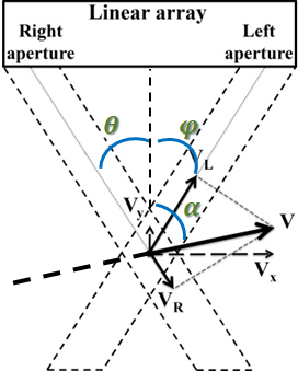
Where θ, φ are ultrasound emission and reception angles, α is the angle between ultrasound probe and target.
After applying the Doppler formula, a vector Doppler formula related to emission and reception angles can be derived:
$$ f_d≅{2\times cos(θ-α)+2\times v\times cos(φ-α)\over c }\times f_0 $$
Rearranging this equation:
$$ v_y (cosθ_m+cosφ_n )+v_x (sinθ_m+sinφ_n )={c\over f_0}\times f_d $$
Using ultrasound information from different angles and the least squares method, the velocity-to-emission-angle relationship in each direction is derived.
$$ Av=u⇒\begin{bmatrix} cos θ_1 + cos φ_1 & sin θ_1 + sin φ_1 \\⋮&⋮\\ cos θ_M + cos φ_N & sin θ_M + sin φ_N \end{bmatrix} \begin{bmatrix}v_z \\ v_x\end{bmatrix} = \begin{bmatrix}u_{11} \\ ⋮ \\ u_{MN}\end{bmatrix} $$
$$v = \begin{bmatrix}v_z \\ v_x\end{bmatrix} = (A^TA)^{-1}A^Tu$$
Results
Projectile Vector Doppler Image of Human Popliteal Vein Valve The image shows local flow direction and velocity changes as blood passes through the venous valve. This type of information can support vascular health research, such as observing flow trends related to thrombosis or varicose veins.